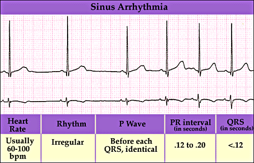
Cardiac dysrhythmia (also known as arrhythmia or irregular heartbeat) is any of a group of conditions in which the electrical activity of the heart is irregular or is
faster or slower than normal. The heartbeat
may be too fast (over 100 beats per minute) or too slow (less than 60 beats per
minute), and may be regular or irregular. A heart beat that is too fast is
called tachycardia
and a heart beat that is too slow is called bradycardia.
Although many arrhythmias are not life-threatening, some can cause cardiac
arrest.
Arrhythmias can occur in the upper chambers of the
heart, (atria), or in the lower chambers of the heart,
(ventricles).
Arrhythmias may occur at any age. Some are barely perceptible, whereas others
can be more dramatic and can even lead to sudden cardiac death.
Signs and symptoms: The term cardiac arrhythmia covers a very large number of
very different conditions.
The most
common symptom of arrhythmia is an abnormal awareness of heartbeat, called palpitations. These may be infrequent, frequent,
or continuous. Some of these arrhythmias are harmless (though distracting for
patients) but many of them predispose to adverse outcomes.
Some
arrhythmias do not cause symptoms, and are not associated with increased
mortality. However, some asymptomatic arrhythmias are associated with
adverse events. Examples include a higher risk of blood clotting within the
heart and a higher risk of insufficient blood being transported to the heart
because of weak heartbeat. Other increased risks are of embolisation and
stroke, heart failure and sudden cardiac death.
If an
arrhythmia results in a heartbeat that is too fast, too slow or too weak to
supply the body's needs, this manifests as a lower blood pressure and may cause
lightheadedness or dizziness, or syncope (fainting).Some types of arrhythmia result in
cardiac arrest, or sudden death.Medical assessment
of the abnormality using an electrocardiogram is one way to diagnose and assess
the risk of any given arrhythmia.
Diagnostic approach: Cardiac dysrhythmias are often first detected by simple but nonspecific
means: auscultation of the heartbeat with a stethoscope, or feeling for peripheral pulses. These cannot usually diagnose specific dysrhythmias, but can give a
general indication of the heart rate and whether it is regular or irregular.
Not all the electrical impulses of the heart produce audible or palpable beats;
in many cardiac arrhythmias, the premature or abnormal beats do not produce an
effective pumping action and are experienced as "skipped" beats.
The simplest specific diagnostic test for
assessment of heart rhythm is the electrocardiogram
(abbreviated ECG or EKG). A Holter
monitor is an EKG recorded over a 24-hour period, to detect dysrhythmias
that may happen briefly and unpredictably throughout the day.
A more advanced study of the heart's electrical
activity can be performed to assess the source of the aberrant heart beats.
This can be accomplished in an Electrophysiology study. A minimally
invasive procedure that uses a catheter to "listen" to the electrical
activity from within the heart, additionally if the source of the arrhythmias
is found, often the abnormal cells can be ablated and the arrhythmia can be
permanently corrected.
Management: The
method of cardiac rhythm management depends firstly on whether or not the
affected person is stable or unstable. Treatments may include physical
maneuvers, medications, electricity conversion, or electro or cryo cautery.
(a)Physical
maneuvers: A number of physical acts can increase
parasympathetic nervous supply to the heart, resulting in blocking of
electrical conduction through the AV node. This can slow down or stop a number
of arrhythmias that originate above or at the AV node (see main article: supraventricular tachycardias).
Parasympathetic nervous supply to the heart is via the vagus nerve, and these
maneuvers are collectively known as vagal maneuvers.
(b)Antiarrhythmic
drugs: There are many classes of antiarrhythmic medications, with different
mechanisms of action and many different individual drugs within these classes.
Although the goal of drug therapy is to prevent arrhythmia, nearly every
antiarrhythmic drug has the potential to act as a pro-arrhythmic, and so must
be carefully selected and used under medical supervision.
(c)Other
drugs: A number of other drugs can be
useful in cardiac arrhythmias. Several groups of drugs slow conduction through
the heart, without actually preventing an arrhythmia. These drugs can be used
to "rate control" a fast rhythm and make it physically tolerable for
the patient. Some arrhythmias promote blood clotting within the heart, and
increase risk of embolus and stroke. Anticoagulant medications
such as warfarin and heparins, and
anti-platelet drugs such as aspirin can reduce the
risk of clotting.
(d)Electricity: Dysrhythmias may also be treated electrically, by
applying a shock across the heart — either externally to the chest wall,
or internally to the heart via implanted electrodes.Cardioversion is either
achieved pharmacologically or via the application of a shock synchronised
to the underlying heartbeat. It is used for treatment of supraventricular
tachycardias. In elective cardioversion, the recipient is usually sedated or
lightly anesthetized for the
procedure. Defibrillation differs in that the shock is not synchronised. It is
needed for the chaotic rhythm of ventricular fibrillation and is also used for
pulseless ventricular tachycardia. Often, more electricity is required for
defibrillation than for cardioversion. In most defibrillation, the recipient
has lost consciousness so there is no need for sedation. Defibrillation or
cardioversion may be accomplished by an implantable cardioverter-defibrillator
(ICD).Electrical treatment of dysrhythmia also includes cardiac
pacing. Temporary pacing may be necessary for reversible
causes of very slow heartbeats, or bradycardia, (for example, from drug
overdose or myocardial infarction). A permanent pacemaker may be placed in situations
where the bradycardia is not expected to recover.
(e)Electrical
cautery: Some
cardiologists further sub-specialise into electrophysiology. In specialised
catheter laboratories, they use fine probes inserted through the blood vessels
to map electrical activity from within the heart. This allows abnormal areas of
conduction to be located very accurately, and subsequently destroyed with heat,
cold, electrical or laser probes. This may be completely curative for some
forms of arrhythmia, but for others, the success rate remains disappointing. AV nodal reentrant tachycardia is often
curable. Atrial fibrillation can also be treated with this technique (e.g.
pulmonary vein isolation), but the results are less reliable.
Reference:
1. Davidson’s Principle and Practice
of Medicine, 21st edition.
2. Wikipedia the free encyclopedia.

মন্তব্যসমূহ
একটি মন্তব্য পোস্ট করুন